Application of dental surgical microscope in the treatment of pulp and periapical diseases
Surgical microscopes have the dual advantages of magnification and illumination, and have been applied in the medical field for more than half a century, achieving certain results. Operating microscopes were widely used and developed in ear surgery in 1940 and in ophthalmic surgery in 1960.
In the field of dentistry, surgical microscopes were applied to dental filling and restoration treatment as early as the early 1960s in Europe. The application of operating microscopes in endodontics truly began in the 1990s, when Italian scholar Pecora first reported the use of dental surgical microscopes in endodontic surgery.
Dentists complete the treatment of pulp and periapical diseases under a dental operating microscope. The dental surgical microscope can magnify the local area, observe finer structures, and provide sufficient light source, allowing dentists to clearly see the structure of the root canal and periapical tissues, and confirm the surgical position. It no longer relies solely on feelings and experience for treatment, thereby reducing the uncertainty of treatment and greatly improving the quality of treatment for pulpal and periapical diseases, enabling some teeth that cannot be preserved by traditional methods to receive comprehensive treatment and preservation.
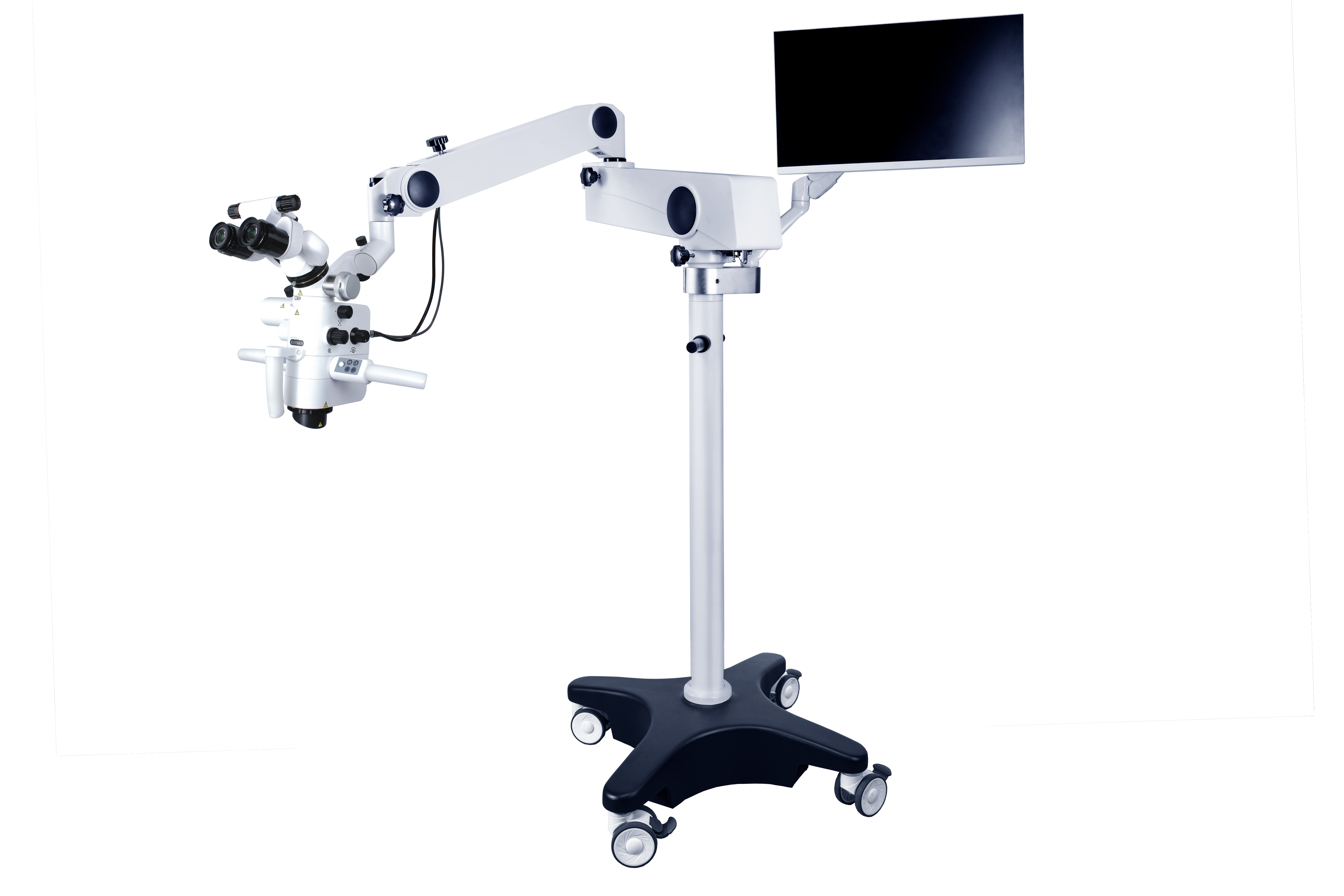
A dental microscope consists of an illumination system, a magnification system, an imaging system, and their accessories. The magnification system is composed of an eyepiece, a tube, an objective lens, a magnification adjuster, etc., which collectively adjust the magnification.
Taking the CORDER ASOM-520-D dental surgical microscope as an example, the magnification of the eyepiece ranges from 10 × to 15 ×, with a commonly used magnification of 12.5X, and the focal length of the objective lens is in the range of 200~500 mm. The magnification changer has two operating modes: electric stepless adjustment and manual continuous magnification adjustment.
The illumination system of the surgical microscope is provided by a fiber optic light source, which provides bright parallel illumination for the field of view and does not produce shadows in the surgical field area. Using binocular lenses, both eyes can be used for observation, reducing fatigue; Obtain a three-dimensional object image. One method to solve the assistant problem is to equip an assistant mirror, which can provide the same clear view as the surgeon, but the cost of equipping an assistant mirror is relatively high. Another method is to install a camera system on the microscope, connect it to a display screen, and allow assistants to watch on the screen. The entire surgical process can also be photographed or recorded to collect medical records for teaching or scientific research.
During the treatment of pulp and periapical diseases, dental surgical microscopes can be used for exploring root canal openings, clearing calcified root canals, repairing root canal wall perforations, examining root canal morphology and cleaning effectiveness, removing broken instruments and broken root canal piles, and performing microsurgical procedures for periapical diseases.
Compared with traditional surgery, the advantages of microsurgery include: precise positioning of the root apex; Traditional surgical resection of bone has a larger range, often greater than or equal to 10mm, while microsurgical bone destruction has a smaller range, less than or equal to 5mm; After using a microscope, the surface morphology of the tooth root can be observed correctly, and the angle of the root cutting slope is less than 10 °, while the angle of the traditional root cutting slope is larger (45 °); Ability to observe the isthmus between root canals at the tip of the root; Be able to accurately prepare and fill root tips. In addition, it can locate the normal anatomical landmarks of the root fracture site and root canal system. The surgical process can be photographed or recorded to collect data for clinical, teaching, or scientific research purposes. It can be considered that dental surgical microscopes have good application value and prospects in the diagnosis, treatment, teaching, and clinical research of dental pulp diseases.

Post time: Dec-19-2024


