How much do you know about surgical microscopes
A surgical microscope is the "eye" of a microsurgery doctor, specifically designed for the surgical environment and typically used to perform microsurgery.
Surgical microscopes are equipped with high-precision optical components, allowing doctors to observe patients' anatomical structures at high magnification and see the most complex details with high resolution and contrast, thereby assisting doctors in performing high-precision surgical operations.
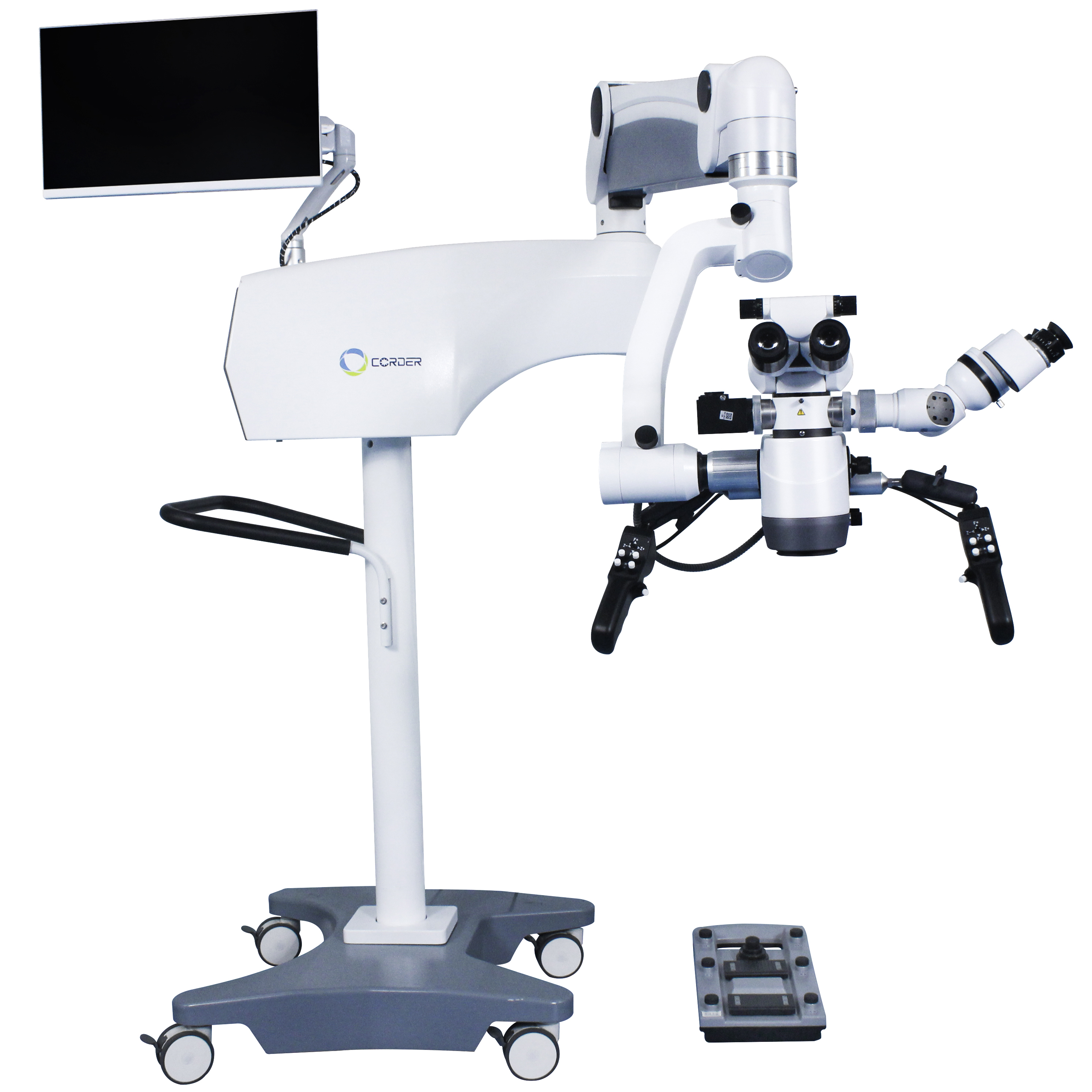
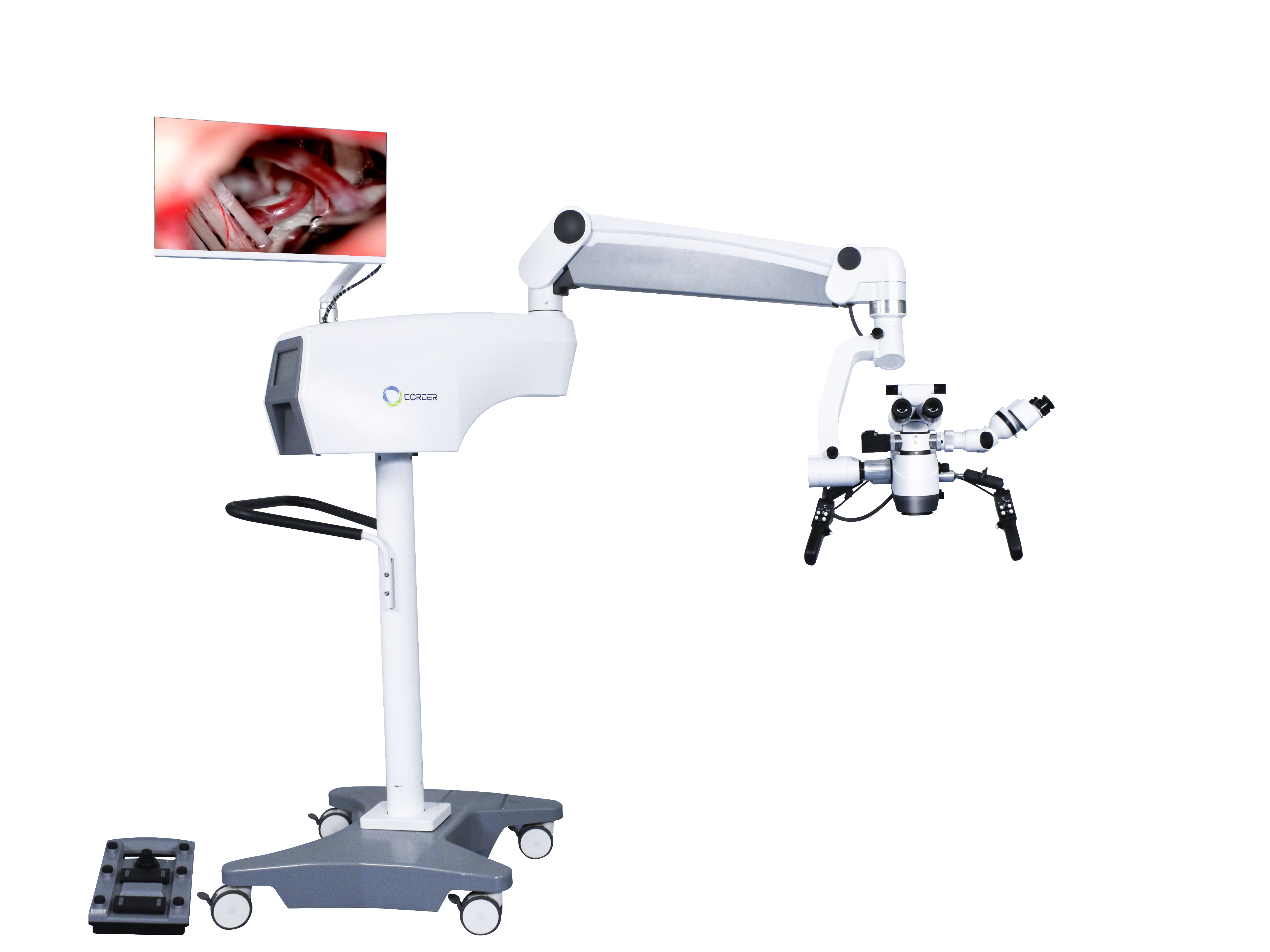
The Operating microscopes mainly consists of five parts: observation system, illumination system, support system, control system, and display system.
Observation system: The observation system mainly consists of an objective lens, a zoom system, a beam splitter, a tube, an eyepiece, etc. It is a key factor affecting the imaging quality of a Medical surgical microscope, including magnification, chromatic aberration correction, and depth of focus (depth of field).
Lighting system: The lighting system mainly consists of main lights, auxiliary lights, optical cables, etc., which is another key factor affecting the imaging quality of Medical surgical microscopes.
Bracket system: The bracket system mainly consists of a base, columns, cross arms, horizontal X-Y movers, etc. The bracket system is the skeleton of the Operating microscope, and it is necessary to ensure the quick and flexible movement of the observation and illumination system to the necessary position.
Control system: The control system mainly consists of a control panel, a control handle, and a control foot pedal. It can not only select operation modes and switch images during surgery through the control panel, but also achieve high-precision micro positioning through the control handle and control foot pedal, as well as control the up, down, left, right focusing of the microscope, the change of magnification, and the adjustment of light brightness.
Display system: mainly composed of cameras, converters, optical structures, and displays.

The development of Professional surgical microscopes has a history of nearly a hundred years. The earliest surgical microscopes can be traced back to the late 19th century, when doctors began using magnifying glasses for surgeries to obtain clearer views. At the beginning of the 20th century, otologist Carl Olof Nylen used a monocular microscope in a surgery for otitis media, opening the door to microsurgery.
In 1953, Zeiss released the world's first commercial surgical microscope OPMI1, which was subsequently applied in ophthalmology, neurosurgery, plastic surgery, and other departments. At the same time, the medical community improved and innovated the optical and mechanical systems of surgical microscopes.
In the late 1970s, after the introduction of electromagnetic switches, the overall structure of Operating microscopes was basically fixed.
In recent years, with the development of high-definition Operating microscopes and digital technology, surgical microscopes have introduced more intraoperative imaging modules and advanced imaging technologies based on their existing performance, such as optical coherence tomography (OCT), fluorescence imaging, and augmented reality (AR), providing doctors with more comprehensive image information.
The binocular surgical microscope generates stereoscopic vision through the difference in binocular vision. In multiple reports, neurosurgeons have listed the lack of stereoscopic visual effects as one of the shortcomings of external mirrors. Even though some scholars believe that three-dimensional stereoscopic perception is not a key factor limiting surgery, it can be overcome through surgical training or by using surgical instruments to move into the temporal dimension of two-dimensional surgical vision to compensate for the lack of three-dimensional spatial perception; However, in complex deep surgeries, two-dimensional endoscopic systems still cannot replace traditional surgical microscopes. Research reports show that the latest 3D endoscope system still cannot completely replace surgical microscopes in key areas of the deep brain during surgery.
The latest 3D endoscope system can provide good stereoscopic vision, but traditional surgical microscopes still have irreplaceable advantages in tissue recognition during deep brain lesion surgery and bleeding. OERTEL and BURKHARDT found in a clinical study of the 3D endoscope system that in a group of 5 brain surgeries and 11 spinal surgeries included in the study, 3 brain surgeries had to abandon the 3D endoscope system and continue using surgical microscopes to complete the surgery during critical steps. The factors that prevented the use of a 3D endoscope system to complete the entire surgical process in these three cases may be multifaceted, including lighting, stereoscopic vision, stent adjustment, and focusing. However, for complex surgeries in the deep brain, surgical microscopes still have certain advantages.

Post time: Dec-05-2024


