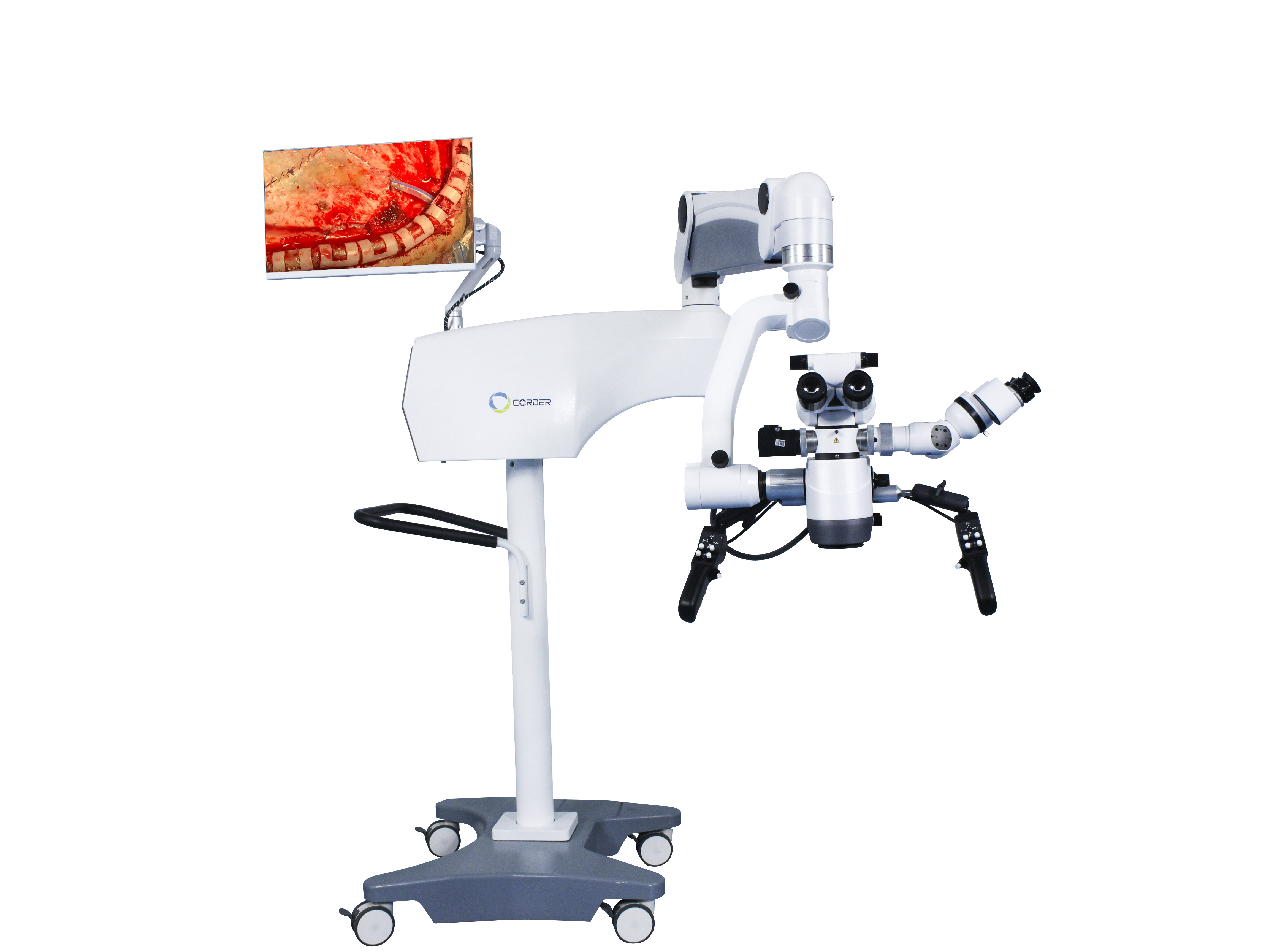
The application history and role of surgical microscopes in neurosurgery
In the history of neurosurgery, the application of surgical microscopes is a groundbreaking symbol, advancing from the traditional neurosurgical era of performing surgery under the naked eye to the modern neurosurgical era of performing surgery under a microscope. Who and when did operating microscopes start to be used in neurosurgery? What role has surgical microscope played in the development of neurosurgery? With the advancement of science and technology, will Operating microscope be replaced by some more advanced equipment? This is a question that every neurosurgeon should be aware of and apply the latest technology and instruments to the field of neurosurgery, promoting the improvement of neurosurgery surgical skills.
1、The History of Microscopy Applications in the Medical Field
In physics, eyeglass lenses are convex lenses with a single structure that have a magnifying effect, and their magnification is limited, known as magnifying glasses. In 1590, two Dutch people installed two convex lens plates inside a slender cylindrical barrel, thus inventing the world's first composite structure magnifying device: the microscope. Afterwards, the structure of the microscope was continuously improved, and the magnification increased continuously. At that time, scientists mainly used this composite microscope to observe the tiny structures of animals and plants, such as the structure of cells. Since the mid to late 19th century, magnifying glasses and microscopes have gradually been applied in the field of medicine. At first, surgeons used eyeglass style magnifying glasses with a single lens structure that could be placed on the bridge of the nose for surgery. In 1876, German doctor Saemisch performed the world's first "microscopic" surgery using a compound eyeglass magnifying glass (the type of surgery is unknown). In 1893, the German company Zeiss invented the binocular microscope, mainly used for experimental observation in medical laboratories, as well as for the observation of corneal and anterior chamber lesions in the field of ophthalmology. In 1921, based on laboratory research on animal inner ear anatomy, Swedish otolaryngologist Nylen used a fixed monocular surgical microscope designed and manufactured by himself to perform chronic otitis media surgery on humans, which was a true microsurgery. One year later, Nylen's superior doctor Hlolmgren introduced a binocular surgical microscope manufactured by Zeiss in the operating room.
The early Operating microscopes had many drawbacks, such as poor mechanical stability, inability to move, illumination of different axes and heating of the objective lens, narrow surgical magnification field, etc. These are all reasons that limit the wider application of surgical microscopes. In the following thirty years, due to the positive interaction between surgeons and microscope manufacturers, the performance of surgical microscopes was continuously improved, and binocular surgical microscopes, roof mounted microscopes, zoom lenses, coaxial light source illumination, electronic or water pressure controlled articulated arms, foot pedal control, and so on were successively developed. In 1953, the German company Zeiss produced a series of specialized surgical microscopes for otology, particularly suitable for surgeries on deep lesions such as the middle ear and temporal bone. While the performance of surgical microscopes continues to improve, the mindset of surgeons is also constantly changing. For example, German doctors Zollner and Wullstein stipulated that surgical microscopes must be used for tympanic membrane shaping surgery. Since the 1950s, ophthalmologists have gradually changed the practice of using only microscopes for ophthalmic examinations and introduced otosurgical microscopes into ophthalmic surgery. Since then, Operating microscope have been widely used in the fields of otology and ophthalmology.
2、Application of surgical microscope in neurosurgery
Due to the particularity of neurosurgery, the application of surgical microscopes in neurosurgery is slightly later than in otology and ophthalmology, and neurosurgeons are actively learning this new technology. At that time, the use of surgical microscopes was mainly in Europe. American ophthalmologist Perrit first introduced surgical microscopes from Europe to the United States in 1946, laying the foundation for American neurosurgeons to use Operating microscopes.
From the perspective of respecting the value of human life, any new technology, equipment, or instruments used for the human body should undergo preliminary animal experiments and technical training for operators. In 1955, American neurosurgeon Malis performed brain surgery on animals using a binocular surgical microscope. Kurze, a neurosurgeon at the University of Southern California in the United States, spent a year learning the surgical techniques of using a microscope in the laboratory after observing ear surgery under a microscope. In August 1957, he successfully performed an acoustic neuroma surgery on a 5-year-old child using an ear surgery microscope, which was the world's first microsurgical surgery. Shortly thereafter, Kurze successfully performed a facial nerve sublingual nerve anastomosis on the child using a surgical microscope, and the child's recovery was excellent. This was the second microsurgical surgery in the world. Afterwards, Kurze used trucks to carry Operating microscopes to various places for microsurgical neurosurgery, and strongly recommended the use of surgical microscopes to other neurosurgeons. Afterwards, Kurze performed cerebral aneurysm clipping surgery using a surgical microscope (unfortunately, he did not publish any articles). With the support of a trigeminal neuralgia patient he treated, he established the world's first micro skull base neurosurgery laboratory in 1961. We should always remember Kurze's contribution to microsurgery and learn from his courage to accept new technologies and ideas. However, until the early 1990s, some neurosurgeons in China did not accept Neurosurgery microscopes for surgery. This was not a problem with the Neurosurgery microscope itself, but a problem with the neurosurgeons' ideological understanding.
In 1958, American neurosurgeon Donaghy established the world's first microsurgery research and training laboratory in Burlington, Vermont. In the early stages, he also encountered confusion and financial difficulties from his superiors. In academia, he always envisioned cutting open cortical blood vessels to directly extract thrombi from patients with cerebral thrombosis. So he collaborated with vascular surgeon Jacobson on animal and clinical research. At that time, under the conditions of the naked eye, only small blood vessels with a diameter of 7-8 millimeters or more could be sutured. In order to achieve end-to-end anastomosis of finer blood vessels, Jacobson first attempted to use a glasses style magnifying glass. Soon after, he recalled using an otolaryngology surgical microscope for surgery when he was a resident physician. So, with the help of Zeiss in Germany, Jacobson designed a dual operator surgical microscope (Diploscope) for vascular anastomosis, which allows two surgeons to perform the surgery simultaneously. After extensive animal experiments, Jacobson published an article on microsurgical anastomosis of dogs and non carotid arteries (1960), with a 100% patency rate of vascular anastomosis. This is a groundbreaking medical paper related to microsurgical neurosurgery and vascular surgery. Jacobson also designed many microsurgical instruments, such as micro scissors, micro needle holders, and micro instrument handles. In 1960, Donaghy successfully performed a cerebral artery incision thrombectomy under a surgical microscope for a patient with cerebral thrombosis. Rhoton from the United States began studying brain anatomy under a microscope in 1967, pioneering a new field of microsurgical anatomy and making significant contributions to the development of microsurgery. Due to the advantages of surgical microscopes and the improvement of microsurgical instruments, more and more surgeons are fond of using surgical microscopes for surgery. And published many related articles on microsurgical procedures.
3、Application of surgical microscope in neurosurgery in China
As a patriotic overseas Chinese in Japan, Professor Du Ziwei donated the first domestic neurosurgical microscope and related microsurgical instruments to the Neurosurgery Department of Suzhou Medical College Affiliated Hospital (now the Neurosurgery Department of Suzhou University Affiliated First Hospital) in 1972. After returning to China, he first performed microsurgical surgeries such as intracranial aneurysms and meningiomas. After learning about the availability of neurosurgical microscopes and microsurgical instruments, Professor Zhao Yadu from the Neurosurgery Department of Beijing Yiwu Hospital visited Professor Du Ziwei from Suzhou Medical College to observe the use of surgical microscopes. Professor Shi Yuquan from Shanghai Huashan Hospital personally visited Professor Du Ziwei's department to observe the microsurgical procedures. As a result, a wave of introduction, learning, and application of Neurosurgery microscopes was sparked in major neurosurgery centers in China, marking the beginning of China's micro neurosurgery.
4、The Effect of Microsurgery Surgery
Due to the use of neurosurgical microscopes, surgeries that cannot be performed with the naked eye become feasible under conditions of magnification of 6-10 times. For example, performing pituitary tumor surgery through the ethmoidal sinus can safely identify and remove pituitary tumors while protecting the normal pituitary gland; Surgery that cannot be performed with the naked eye can become better surgeries, such as brainstem tumors and spinal cord intramedullary tumors. Academician Wang Zhongcheng had a mortality rate of 10.7% for cerebral aneurysm surgery before using a neurosurgery microscope. After using a microscope in 1978, the mortality rate decreased to 3.2%. The mortality rate of cerebral arteriovenous malformation surgery without the use of a surgical microscope was 6.2%, and after 1984, with the use of a neurosurgery microscopes, the mortality rate decreased to 1.6%. The use of neurosurgery microscope allows pituitary tumors to be treated through a minimally invasive transnasal transsphenoidal approach without the need for craniotomy, reducing the surgical mortality rate from 4.7% to 0.9%. The achievement of these results is impossible under traditional gross eye surgery, so surgical microscopes are a symbol of modern neurosurgery and have become one of the indispensable and irreplaceable surgical equipment in modern neurosurgery.

Post time: Dec-09-2024


